Faculty
Gerard Ahern

Gerard Ahern
Associate Professor, Pharmacology and Physiology
gpa3@georgetown.edu
Education: B.Sc. (Hons) 1990, LL. B. University of Canterbury NZ, 1991, Ph.D. Australian National University, 1996
Current Research: How do cells detect changes in the external or extracellular environment? We are interested in the fundamental mechanisms that allow cells to sense diverse chemical or physical stimuli. Our focus is a class of membrane ion channels called “Transient Receptor Potential” (TRP) channels. We also explore novel ligand signaling at G-protein coupled receptors both in neurons and immune cells. We use a combination of electrophysiological, cell imaging, genetic and biochemical techniques, and where possible, appropriate animal models.
Tinatin I. Brelidze
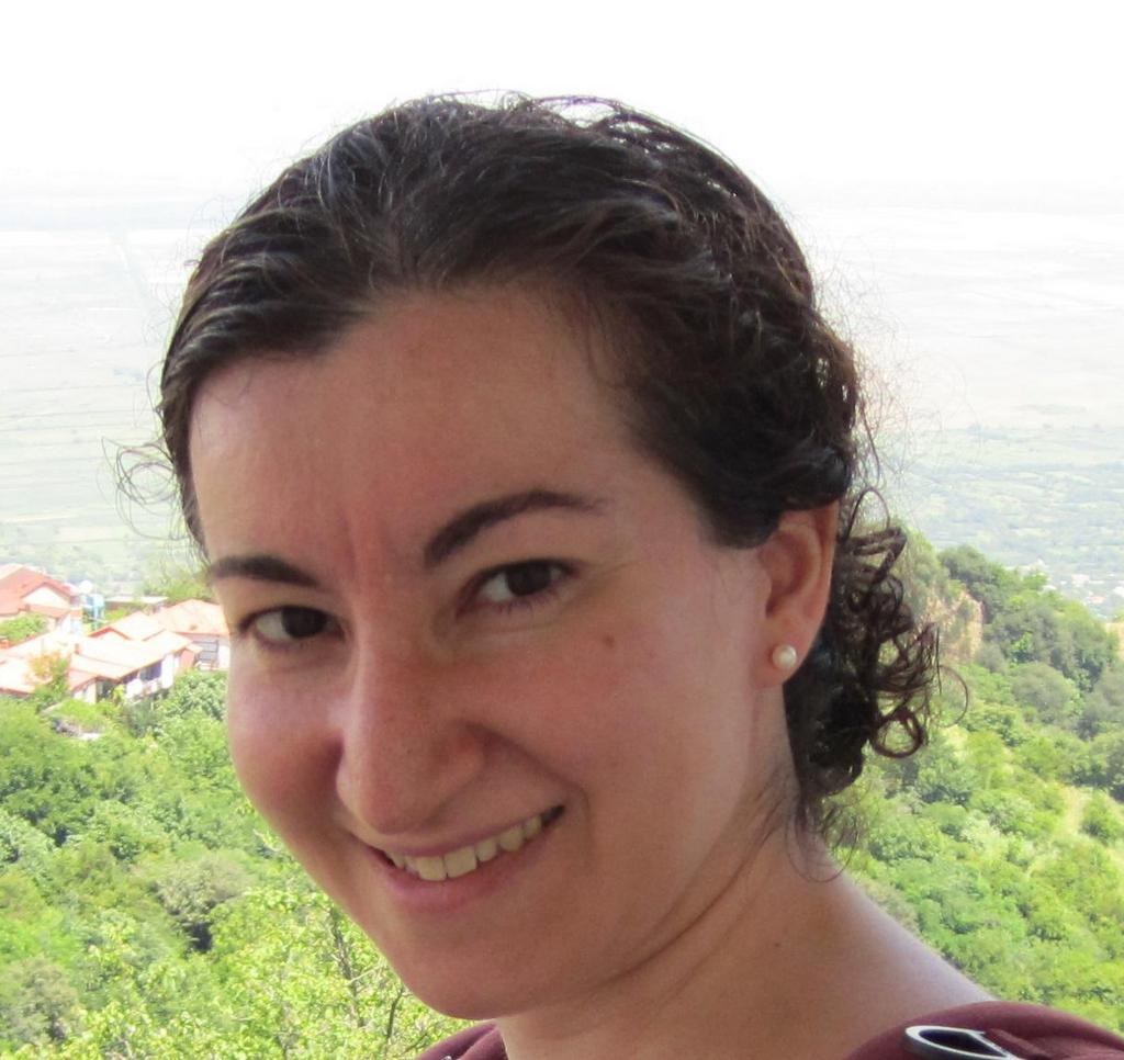
Tinatin I. Brelidze
Associate Professor, Pharmacology & Physiology
tib5@georgetown.edu
Education: Diploma in Physics (Hons), Tbilisi State University (Georgia), 1996; Ph.D. Physiology & Biophysics, University of Miami, 2003
Current Research: Ion channels are guardians of membrane potential and are essential for the physiological function of every living cell. Abnormalities in ion channel opening and closing (gating) or expression pattern are often linked to inherited or acquired diseases. Our research interests are focused on understanding the mechanisms of ion channel gating. To uncover the novel mechanisms of ion channel gating we use a combination of electrophysiology, X-ray crystallography and fluorescence based methods.
Casey Brown

Casey Brown
Assistant Professor, Psychology
casey.brown@georgetown.edu
Education: University of Virginia: B.A. (High Distinction), Cognitive Science & Psychology, 2011; Ph.D. Clinical Science, University of California, Berkeley, 2020
Current Research: Emotion, empathy, social emotion regulation, depression/mental & physical health, socioemotional functioning in neurodegenerative and neuropsychiatric disorders, aging and dementia, caregiving, time-horizons, stress, nonverbal behavior, psychophysiology & interoception, neuroimaging.
Mark P. Burns

Mark P. Burns
Professor, Neuroscience
Laboratory for Brain Injury and Dementia
mpb37@georgetown.edu
Education: National University of Ireland, Galway: B.Sc. (Hons), Physiology, 1997; Ph.D. Pharmacology, 2000
Current Research: The Laboratory for Brain Injury and Dementia (LBID) at Georgetown University studies the acute activation of pathways involved in chronic neurodegenerative diseases such as Alzheimer’s disease after traumatic brain injury (TBI) Our aim is to understand if these pathways are playing a role in acute cell death after TBI, and to to understand if the activation of these pathways are involved in development of dementias, such as Alzheimer’s disease and chronic traumatic encephalopathy (CTE). We use contusion and concussive brain trauma models in the LBID, and the lab has capability to perform the surgical, behavioral and biochemical aspects of this research. We take advantage of the outstanding core imaging facilities available at Georgetown University, including a small animal imaging laboratory with a 7Tesla magnetic resonance imager, confocal microscopy, stereology, fluorescent and standard microscopy.
Isaac Cervantes-Sandoval

Isaac Cervantes-Sandoval
Assistant Professor, Department of Biology
Cervantes Lab Website
ic400@georgetown.edu
Education: B.S. Instituto Politecnico Nacional (IPN)-Mexico; M.S., Ph.D. Centro de Investigación y Estudios Avanzados (CINVESTAV)–México
Current research: Molecular and cellular mechanisms of learning and memory; forgetting memories using the fruit fly as an animal model. Our lab is trying to understand how memories are encoded in the brain and how they are forgotten. For this, we use the relatively well-studied and simple Drosophila brain, a biological model that offers tremendous advantages in the study of neuroscience.
Thomas M. Coate

Thomas M. Coate
Associate Professor, Biology
Coate Lab
tmc91@georgetown.edu
Education: B.S. University of Puget Sound; Ph.D. Oregon Health & Science University
Current Research: The long-term goal of the research in the Coate laboratory will be to define the signaling mechanisms underlying neural development within sensory systems and how synaptic connections can be reestablished in cases of damage or disease. In the field of developmental neuroscience, we are now at an exciting time where we can take multi-faceted approaches to understand (with excellent temporal and spatial resolution) the mechanisms by which precise cell types coordinate appropriate axon guidance decisions and synapse formation. In our research, we aim to understand the mechanisms by which spiral ganglion neurons (SGNs) make functional connections with mechanosensory hair cells in the mouse cochlea. We are currently addressing how secreted Semaphorins, which activate Neuropilin/Plexin co-receptors, control SGN axon guidance decisions in the cochlear sensory epithelium. We are also investigating how the transcription factor Pou3f4 controls the expression of axon guidance factors in the developing cochlea (such as ephrins and Eph receptors) and how those factors control cochlear innervation. The cochlea provides an excellent model for discovering how circuits assemble within a complex organ system, as it is composed of an array of cell types and structures precisely arranged to detect a range of sound frequencies.
Katherine Conant
Katherine Conant
Professor, Neuroscience
kec84@georgetown.edu
Education: A.B. Cornell University, Biochemistry; M.D. Boston University
Current Research: Matrix metalloproteinases (MMPs) are a family of zinc-dependent endopeptidases that are released in a neuronal activity-dependent manner. MMP expression and activity can also be dramatically upregulated with an injury. Many studies have therefore focused on their role in pathology. Less is known about the role of MMPs in normal CNS physiology, and whether critical physiological processes might be disrupted when MMP levels are pathologically elevated. While recent studies suggest that MMPs play a role in learning and memory, the mechanisms by which they do so are not well understood. Similarly, how excessive MMP activity might contribute to synaptic injury is not clear. Our work is focused on specific mechanisms by which MMPs can influence neuronal and synaptic structure and function. Our focus is on cleavage of synaptic adhesion molecules and thrombin type G protein coupled receptors.
Andrew DeMarco
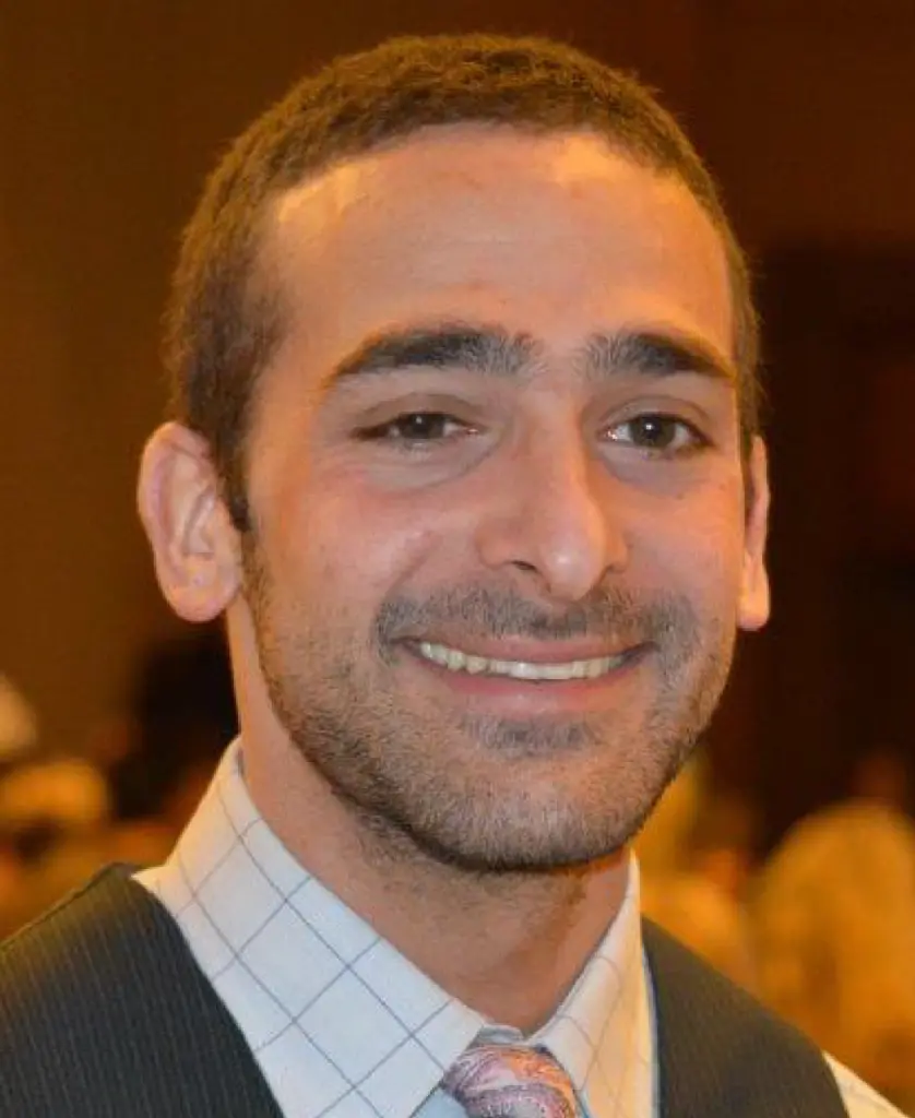
Andrew DeMarco
Assistant Professor, Department of Rehabilitation Medicine
DeMarco Advanced Research in Neurorehabilitation (DARN) Lab
andrew.demarco@georgetown.edu
Education: Ph.D., CCC-SLP, University of Arizona, 2016
Current Research: Dr. DeMarco is a cognitive neuroscientist and speech-language pathologist whose research investigates the neural and cognitive architecture of speech and language, how this circuitry breaks down after brain injury such as a stroke, and how this information can be used to ultimately inform clinical decision-making in neurorehabilitation to optimize patient outcomes. The lab studies both healthy individuals and individuals with acquired language disorders, primarily stroke survivors with aphasia. We currently utilize a variety of techniques including structural, diffusion, and functional MRI, plus EEG and detailed neuropsychological testing. Our work emphasizes technical innovation and novel analytical approaches with an eye toward clinical translation of imaging biomarkers. We also focus on big data approaches by leveraging our close partnership with the MedStar hospital network.
Rhonda Dzakpasu

Rhonda Dzakpasu
Associate Professor, Physics and Pharmacology
Neural Dynamics Lab
dzakpasu@physics.georgetown.edu
Education: Ph.D. University of Michigan, 2003
Current Research: We use arrays of extracellular multi-electrodes to record and stimulate electrical activity from cultured neural circuits as well as from acute neural slices. We modulate network rhythmicity by manipulating the balance between excitation and inhibition to investigate the principles by which neurons interact. What is the causal role of emergent coherent activity for neuronal communication?
Guinevere Eden

Guinevere Eden
Professor, Pediatrics
Center for the Study of Learning
edeng@georgetown.edu
Education: B.Sc. University College London, Physiology, 1989; D.Phil. Oxford University, Physiology, 1993.
Current Research: Dr. Eden’s research has focused on the application of functional neuroimaging techniques to study the neural basis of reading and how it may be altered in individuals with developmental disorders or altered early sensory experience. Further, she and her colleagues are researching how reading is impacted by instructions or mode of communication and are utilizing functional MRI to study the neurobiological correlates of reading remediation.
Rebekah Evans

Rebekah Evans
Assistant Professor, Neuroscience
Evans Lab Website
re285@georgetown.edu
Education: B.A. St John’s College 2004, M.Ed. George Mason University 2006, Ph.D. Neuroscience George Mason University 2013
Current Research: The Evans Lab studies neural dynamics and functional connectivity in brain structures that degenerate in Parkinson’s Disease. Dr. Evans’ lab uses systems, cellular, and computational neuroscience approaches to test how circuit alterations within the brain can protect vulnerable neurons from neurodegeneration. To probe circuitry, the Evans lab uses two-photon calcium imaging with simultaneous optogenetics to image neural activity under highly specific stimulation conditions in active brain slices. To further dissect these circuits, in vivo optogenetics and calcium imaging approaches are employed to control and record neural activity during animal behavior. At the cellular level, the Evans lab images dendrites and dendritic spines during synaptic activity to understand how specific inputs modulate information processing within a single cell.
Patrick A. Forcelli

Patrick A. Forcelli
Professor, Pharmacology & Physiology
Laboratory of Molecules, Circuits and Behavior
paf22@georgetown.edu
Education: Ph.D. Georgetown University, Neuroscience, 2011
Current Research: 1. The vulnerability of the developing brain to drug-induced damage – Anti-seizure medications (also called an anticonvulsant or antiepileptic drugs) are the mainstay of treatment for epilepsy. However, the use of these medications in special populations (e.g., pregnant women with epilepsy, neonates with seizures) poses a challenge due to the exquisite sensitivity of the developing brain to perturbations of neural activity. Many commonly utilized anti-seizure drugs induce apoptosis in the neonatal rat brain, disrupt functional and morphological synaptic development, and alter behavioral function. We are continuing to examine this class of drugs to characterize effects on brain development, identify mechanisms of toxicity, and search for therapeutic approaches to minimize long-term effects. We employ a combination of histological, biochemical, physiological, behavioral, and imaging approaches to address these questions.
2. Hippocampal and basal ganglia circuits in seizure progression. The same neural circuits through which seizures propagate are vital participants in the normal cognitive and emotional function. We are examining hippocampal and basal ganglia contributions to both normal behaviors and to seizure propagation. To determine the role of these circuits we are using a combination of lesions, focal pharmacological (in rodents and primates), electrical, and state of the art pharmacogenetic and optogenetic methods. These techniques are used to perturb circuit function during behavioral tasks as well as in animal models of seizures/epilepsy. Finally, we are also assessing the ability of shRNA-mediated knockdown of select targets within these circuits to delay or prevent the development of epilepsy.
Rhonda B. Friedman

Rhonda B. Friedman
Professor, Neurology
Center for Aphasia Research and Rehabilitation
RFriedman@georgetown.edu
Education: B.A. University of Pennsylvania, 1974; Ph.D. MIT, 1978
Current Research: Dr. Friedman’s research focuses on how language is processed in the normal brain; how language breaks down in a brain damaged by stroke, head injury, or dementia; how the brain recovers language function after injury; and how the recovery process can be aided by non-pharmaceutical therapies. Research focuses particularly on semantic memory, naming, and reading. Techniques employed include behavioral studies; treatment studies; ERP; eye-tracking; and imaging studies. Current studies focus on learning paradigms in rehabilitation, and prophylaxis of cognitive decline in dementia.
Nady Golestaneh

Nady Golestaneh
Associate Professor, Ophthalmology, Neurology, Biochemistry
Golestaneh Lab
ncg8@georgetown.edu
Education: B.S. University of Paris VII, Jussieu; M.S., Ph.D., University of Paris VI, Pierre et Marie Curie, Paris, France
Current Research: Dr. Golestaneh’s lab is investigating how aging mechanisms affect the cells and induce diseases such as age-related macular degeneration (AMD). Using patient-specific induced pluripotent stem (iPS) cells Dr. Golestaneh is studying the cellular and molecular mechanisms that induce the AMD and is developing ways to stop this disease from occurring. Another focus of the lab is developing new methods of autologous cell-based therapy for AMD by generating patient-specific stem cell-derived neuroretinal cells.
Adam Green

Adam Green
Associate Professor, Psychology
Georgetown Laboratory for Relational Cognition
aeg58@Georgetown.edu
Education: Ph.D. Dartmouth College, 2007.
Current Research: My motivating interest is in human creative intelligence and especially in understanding how neural processes constitute our best ideas. Adam’s work focuses on reasoning that reveals meaningful connections between seemingly distant concepts, often in the form of analogies.
Anna Greenwald
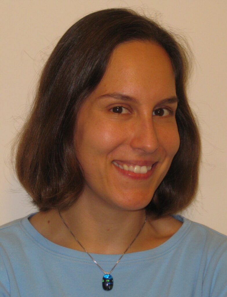
Anna Greenwald
Research Assistant Professor
Center for Brain Plasticity and Recovery
as2266@georgetown.edu
Education: Ph.D. University of Giessen, Germany, Psychology, 2009
Current Research: Dr. Greenwald’s research employs behavioral and magnetic resonance imaging (MRI) methods to investigate specific cognitive functions (e.g., visuo-spatial cognition, language, attention) and their functional neuroanatomy. She works with patients who have acquired brain injury at different points in life (e.g., stroke during the perinatal period, during childhood, or in adulthood) as well as neurologically healthy controls. Her long-term goal is to improve assessment tools and rehabilitation options for patients with cognitive impairments.
Haiyan He
Haiyan He
Assistant Professor
He Lab Website
haiyan.he@georgetown.edu
Education: B.S. Shanghai Jiaotong University, China; M.S. Peking University, China; Ph.D. University of Maryland, College Park, M.D.
Current Research: The long-term goal of He lab is to unravel the molecular and cellular mechanisms underlying the intricate balance between plasticity and stability of neural circuits in both early embryonic development and adulthood, with a special focus on inhibitory neurons. We use the albino Xenopus laevis (tadpole) as our animal model. The central nervous system of the tadpole is amenable to a wide variety of manipulations from molecular to circuit level. It provides a unique in vivo vertebrate system to study the basic laws governing the organization and formation of nascent neural circuits, where plasticity and stability are both pivotal for the survival of the animal. We use a number of techniques in the lab, including time-lapse in vivo imaging of neuronal structure and function and using bio-orthogonal non-canonical amino acid (BONCAT) to tag activity-induced newly synthesized proteins, as well as behavioral evaluation of visual functions. Current projects include investigating how excitatory and inhibitory synaptic inputs are coordinated in response to experience changes and examining functional roles of the degradation of newly-synthesized protein in maintaining neuronal homeostasis.
Jeffrey K. Huang

Jeffrey K. Huang
Associate Professor, Biology
Huang Laboratory
jh1659@georgetown.edu
Education: B.A. Washington University; Ph.D. Mount Sinai School of Medicine of NYU
Current Research: My lab is interested in the biology and pathology of glial cells. We focus on oligodendrocytes, a type of glia, whose cellular processes engage with and enwrap CNS axons, and form the lipid-rich myelin membranes required for rapid, saltatory conduction. Myelin destruction in diseases such as multiple sclerosis impairs axonal conduction and results in progressive axonal degeneration. We are currently investigating the mechanisms by which oligodendrocytes interact and communicate with axons, and how their interactions might promote axonal integrity and survival. We are also investigating the mechanism of myelin regeneration, with a focus on how oligodendrocytes regenerate from endogenous neural progenitor cells to replace myelin during homeostatic turnover or after demyelination. We use primary oligodendrocyte/neuron co-cultures, transgenic mice, and models of experimental CNS injury and demyelination, combined with molecular biology and imaging tools to address these questions.
Xiong Jiang
Xiong Jiang
Associate Professor, Neuroscience
Cognitive Neuroimaging Laboratory
Cognitive Neuroimaging Laboratory Website
xj9@georgetown.edu
Education: Ph.D. The Catholic University of America, Experimental Psychology, 2005
Current Research: Magnetic resonance imaging (MRI) has a strong potential to serve as non-invasive research and clinical tool of neurological disorders. Our lab has been working to develop and validate novel and advanced MRI techniques that are sensitive to subtle neuronal injury at the early stages of the disease, with a focus on functional MRI (fMRI). The ultimate goal is to develop multimodal MRI biomarkers that can effectively detect and accurately quantify disease progression, even in the absence of behavioral symptoms, with the potential to guide and evaluate early interventional therapies.
Jagmeet Kanwal
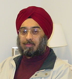
Jagmeet Kanwal
Associate Professor, Neurology, Neuroscience, and Psychology
Complex Adaptive Systems Neuroscience
kanwalj@georgetown.edu
Education: Ph.D. LSU-Baton Rouge, Physiology and Zoology, 1986
Current Research: My lab is equipped to use sounds and computational models to probe neural circuits so that we can understand how the brain works, what its design features are, and how neurons compute. We tackle these questions using a multiplicity of techniques, organisms, and approaches to obtain neurometrics across a wide range of spatial, spectral, and temporal scales. Topics of interest: 1. Sensory coding and computational plasticity: how are species-specific sounds encoded/decoded within the cortex and amygdala? Solution of this nontrivial problem may inform us about the origins and mechanisms for speech and music perception and facilitate design of neuroprosthetic devices. 2. Cortico-cortical and cortico-limbic interactions: We study the role of functional connectivity, oscillations and single-unit responses. 3. Learning, Memory, and Distress: The brain exhibits multistate and multisite adaptive plasticity for decision-making. How do anxiety, traumatic stress, and autism spectrum disorders emerge?
Ken Kellar

Ken Kellar
Professor, Pharmacology and Physiology
Kellar Lab
kellark@georgetown.edu
Education: Ph.D. Ohio State University, Pharmacology, 1974
Current Research: My laboratory studies the subunit composition, pharmacological properties and regulation of neuronal nicotinic receptors. We are particularly interested in understanding the importance of desensitization of these receptors and the mechanisms by which chronic exposure to nicotine increases these receptors in brain.
Ian Lyons

Ian Lyons
Assistant Professor, Psychology
Math Brain Lab
ian.lyons@georgetown.edu
Education: Ph.D. University of Chicago, Psychology (Cognitive), 2012
Current Research: In our lab, we use concepts and tools from developmental psychology, cognitive science, and cognitive neuroscience to understand how humans acquire, use and teach numerical and mathematical concepts. Research interests include numerical cognition; mathematical thinking and learning; math anxiety; math education – social, cognitive and emotional factors; acquisition of symbolic systems – numerical symbol representation; cognitive development; cognitive neuroscience.
Kathy Maguire-Zeiss

Kathy Maguire-Zeiss
Professor & Chair, Neuroscience
Maguire-Zeiss Lab
km445@georgetown.edu
Education: B.S. Albright College, 1981; Ph.D. Pennsylvania State University College of Medicine, 1987
Current Research: My laboratory is focused on understanding the mechanisms involved in age-related progressive neurodegenerative diseases. Specifically, we are investigating the role of increased oxidative stress, protein misfolding and inflammation using mouse transgenic and toxicant models as well as cell culture technologies. Our work includes studies with the Parkinson’s disease protein, α-synuclein, and how it increases oxidative stress and proinflammatory molecules, as well as, the role of inflammation in HIV-associated neurocognitive disorder. We hope our studies will help us to better understand how inflammation is involved early in PD and HAND.
Ludise Malkova
Ludise Malkova
Professor, Pharmacology
malkoval@georgetown.edu
Education: B.A., M.A. Charles University Prague, Czech Republic; Ph.D. Czechoslovak Academy Sciences, Prague, Czech Republic, 1986
Current Research: Neural substrates of social and emotional behavior; the role of the amygdala and orbitofrontal cortex in processing reward; medial temporal lobe structures (hippocampus, perirhinal cortex) and cognitive functions (object recognition and spatial memory); amygdala and midbrain (superior colliculus) interactions; autism, PTSD; reversible pharmacological manipulations of discrete brain structures, systemic drug effects.
Abigail Marsh

Dr. Abigail Marsh,
Abigail Marsh
Professor, Psychology
Laboratory on Social & Affective Neuroscience
aam72@georgetown.edu
Education: Ph.D. Harvard University, Social Psychology, 2004
Current Research: How do people understand what others think and feel? How does that relate to what they think and feel themselves? What causes people to want to help or harm others? These are the questions that underlie research on empathy and mentalizing. Research in the lab is aimed at understanding aspects of human social interactions, emotional functioning, and empathy using cognitive neuroscience methods. We focus in particular on emotion and on nonverbal communication. Our research includes studies with adolescents and adults, incorporating neuroimaging, cognitive and behavioral testing, and psychopharmacological techniques.
Andrei Medvedev

Andrei Medvedev
Associate Professor, Neurology
Medvedev Lab
am236@georgetown.edu
Education: Ph.D. Russian Academy of Sciences, Moscow, Neurophysiology, 1989
Current Research: Dr. Medvedev’s expertise includes systems electrophysiology, signal analysis and neural network modeling. His research interests focus on neural mechanisms of cognitive processes in normal and neurological pathology. His current research combines a new technology of noninvasive near-infrared (NIR) optical functional imaging of the brain with a more traditional electrophysiological analysis of on-going and event-related electrical activity (EEG, ERP, evoked and induced gamma-band oscillations).
Italo Mocchetti

Italo Mocchetti
Professor, Neuroscience
moccheti@georgetown.edu
Education: Ph.D. University of Milan, Italy, 1982
Current Research: Neurotrophic factors influence axon and dendrite growth, synaptic plasticity and neurogenesis, and the interaction of neurons with glial cells. They play critical roles in preventing neurological diseases including Alzheimer’s disease, Parkinson’s disease, Huntington’s disease, stroke and epilepsy, but they can also promote neuronal apoptosis. Our laboratory has recently demonstrated that the neurotrophin brain-derived neurotrophic factor (BDNF) modulates the expression and function of chemokine receptors that contribute to AIDS dementia complex. We envision this research as catalyzing important new efforts to translate the basic science of the neurotrophins into effective new treatments for neurodegenerative diseases. In addition, we study the effect of opioids on the neuronal survival.
Charbel Moussa

Charbel Moussa
Associate Professor, Neurology
The Laboratory for Dementia and Parkinsonism
cem46@georgetown.edu
Education: M.B. (M.D. equivalent), Ph.D. University of Sydney, Australia, 2002
Current Research: Our laboratory evaluates potential therapeutic drugs for neurodegenerative diseases in pre-clinical models and clinical trials. We investigate cellular, biochemical and pathological mechanisms that underlie neurodegenerative diseases with dementia and Parkinsonism, including Alzheimers disease (AD) and other dementias, and Parkinsons disease (PD) and related movement disorders. We also investigate the neuro-pathological and biochemical pathways involved in the spectrum of motor neuron disease (MND) with fronto-temporal dementia (FTD).
Elissa L. Newport
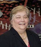
Elissa L. Newport
Professor of Neurology, Director of CBPR, Neurology
Center for Brain Plasticity and Recovery
eln10@georgetown.edu
Education: Ph.D. University of Pennsylvania, 1975
Current Research: Dr. Newport’s research examines language acquisition and other types of implicit pattern learning. We focus on young children, asking how they acquire spoken or sign languages, even when they may have little consistent or complex linguistic input. Our studies include fieldwork on young, developing sign languages around the world, and also experiments in the lab, investigating how children and adults learn miniature artificial languages that reproduce specific properties of natural languages and their acquisition circumstances. We have shown that children expand languages as they learn them, making them more consistent and shifting them toward universal principles of language structure. We also study how children and adults acquire or recover language after left hemisphere strokes. This research investigates the mechanisms underlying language acquisition and language reorganization after brain injury, with a particular interest in developmental plasticity.
Alexey Ostroumov
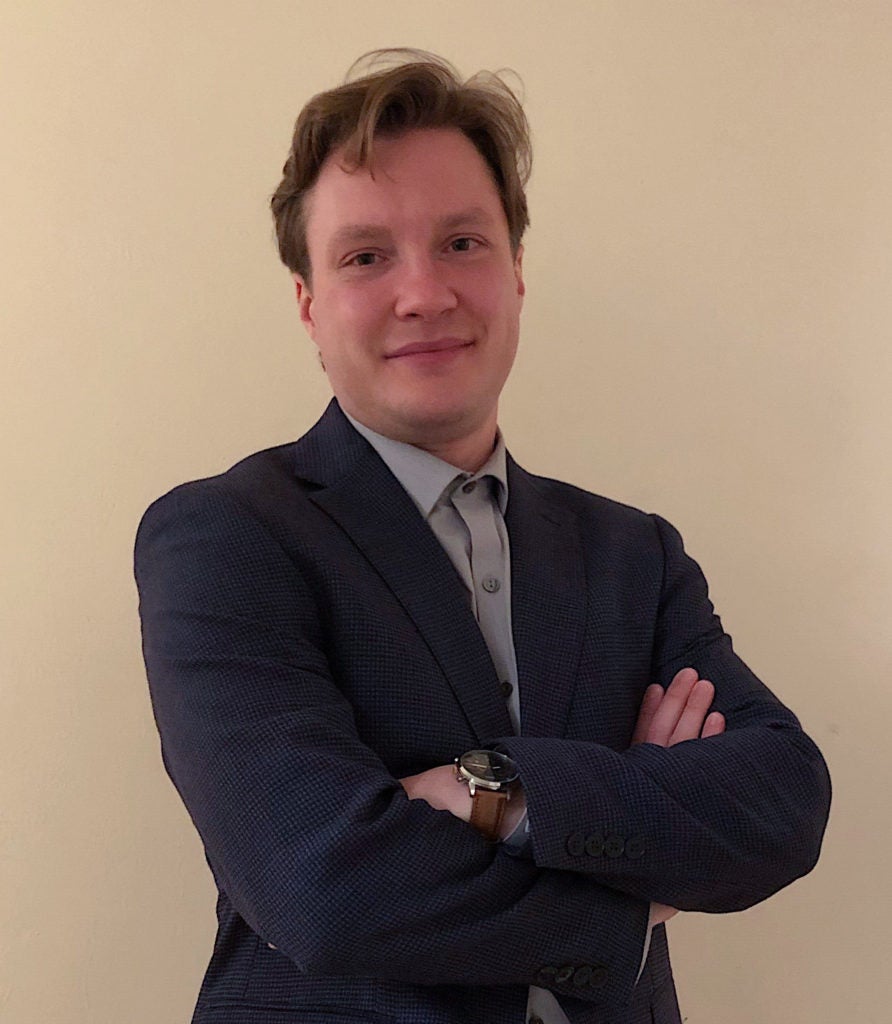
Alexey Ostroumov
Assistant Professor, Pharmacology and Physiology
Ostroumov Lab Website
ao711@georgetown.edu
Education: Ph.D. International School for Advanced Studies (SISSA, Italy), 2010
Current research: An organism adapts its behavior through brain circuitry remodeling, which results in part from experience-dependent synaptic plasticity. Traumatic life experiences, chronic stress, and addictive drugs all dysregulate adaptive mechanisms, producing a variety of aberrant behaviors associated with neuropsychiatric disorders, such as depression, anxiety and addiction. The main goal of my laboratory is to provide mechanistic insights into the aberrant behaviors and thereby contribute to the development of novel and effective treatments of neuropsychiatric disorders. Specifically, we seek to understand how experience modifies synapses and neural circuitry within the mesocorticolimbic dopamine system and how these changes impact reward processing, motivation, and executive function. To this end, we apply a multi-disciplinary approach, combining pharmacological and electrophysiological techniques with behavior and state-of-the-art viral and optogenetic tools.
Daniel Pak

Daniel Pak
Professor, Pharmacology and Physiology
Molecular Neurobiology of Memory [mNeMe]
dtp6@georgetown.edu
Education: B.A. Harvard University, 1991; Ph.D. University of California at Berkeley, 1996
Current Research: My laboratory is interested in the molecular changes that occur at CNS synapses in response to experience. We utilize a combination of approaches ranging from molecular biology and biochemistry to cell biology, imaging, and mouse genetics to address three major questions: 1) how do neurons encode long-term storage of information? 2) how do neurons maintain stability of function by homeostatic synaptic plasticity mechanisms? and 3) how does failure of these mechanisms contribute to neurological and neurodegenerative disorders?
Josef P. Rauschecker

Josef P. Rauschecker
Professor, Neuroscience
Laboratory for Integrative Neuroscience and Cognition
rauschej@georgetown.edu
Education: Ph.D. Munich Institute of Technology, 1980; D.Sc. Tubingen University, Neurophysiology, 1985
Current Research: Dr. Rauschecker is interested in the functional organization and plasticity of the cerebral cortex. He is especially interested in how perception, memory, and language are implemented by the brain. His laboratory is one of only a handful in the country engaged in the neurophysiology of auditory cortex in nonhuman primates. In parallel studies, he is using functional magnetic resonance imaging (fMRI) in humans for the study of the neural basis of language, music and other higher auditory processing. His laboratory is also interested in the effects of sensory deprivation during brain development, relating to the question of how the brain of individuals with early blindness or deafness gets reorganized.
G. William Rebeck

G. William Rebeck
Professor, Department of Neuroscience
Rebeck Lab
gwr2@georgetown.edu
Education: A.B. Cornell University, 1982; Ph.D. Harvard University, 1991
Current Research: I have been studying the effect of APOE on Alzheimer’s disease pathogenesis since 1993. The APOE4 allele is the strongest genetic risk factor for Alzheimer’s disease. We study the role of the APOE in neuronal signaling and glial activation, cell culture systems and APOE knock-in mice, which express the human APOE alleles under the endogenous mouse APOE promoter. We have found that the APOE4 isoform is altered in its post-translational modifications in the CNS compared to other APOE isoforms. We have also found that the APOE4 mice have reduced neuronal complexity and increased sensitivity to glial activation. These effects of APOE occur in the absence of Alzheimer’s disease pathological changes, suggesting that APOE modifies normal brain functions. We have found that APOE4 mice are also more susceptible to cognitive impairment after chemotherapy. Altering APOE regulation and modification in normal brains presents opportunities to prevent cognitive impairments due to Alzheimer’s disease and chemotherapy.
Maximilian Riesenhuber

Maximilian Riesenhuber
Professor, Neuroscience
Laboratory for Computational Cognitive Neuroscience
mr287@georgetown.edu
Education: Ph.D. MIT, 2000
Current Research: The lab investigates the computational mechanisms underlying human object recognition as a gateway to understanding the neural bases of intelligent behavior. We combine computational models with human behavioral, functional magnetic resonance imaging (fMRI) and electroencephalographic (EEG) data. This comprehensive approach addresses one of the major challenges in cognitive neuroscience today: the necessity to combine data from a range of experimental levels in order to develop a rigorous and predictive model of human brain function that quantitatively and mechanistically links neurons to behavior. This computational grounding of behavior in processing at the neuronal level is of key interest for the investigation of the neural bases of cognitive and behavioral impairments in complex mental disorders, to pave the way for neuroscience-based therapies. In addition, understanding the neural mechanisms underlying object recognition in the brain is of significant interest for Artificial Intelligence, as the capabilities of pattern recognition systems in engineering (e.g., in machine vision or speech recognition) still lag far behind that of their human counterparts in terms of robustness, flexibility, and the ability to learn from few exemplars.
Kathryn Sandberg

Kathryn Sandberg
Professor, Nephrology & Hypertension, Dept. of Medicine; Director, CSD
Center for the Study of Sex Differences
sandberg@georgetown.edu
Education: Ph.D. University of Maryland, Baltimore, Biochemistry, 1987
Current Research: Dr. Sandberg’s laboratory is investigating the structural analysis and regulation of G protein-coupled receptors in neuronal physiology and pathophysiology. In particular, her research focuses on the receptors for angiotensin II, corticotropin releasing hormone, gonadotropin releasing hormone, vasopressin and neuropeptide Y and their role in neuronal and cardiovascular physiology and pathophysiology. A second major aim of this laboratory is to study mechanisms of G protein-coupled receptor gene regulation. Dr. Sandberg is especially interested in understanding mechanisms of gene control at the posttransciptional level.
Elena Silva

Elena Silva
Professor, Biology
Casey Lab
emc26@georgetown.edu
Education: Ph.D. Stanford University, Biology, 1996
Current Research: Our goal is to define the gene network that controls the induction and differentiation of the central nervous system. The CNS is derived from the ectoderm which can develop into either epidermal or neural tissue. Neural tissue then differentiates into either neurons or glial cells. Many of the signal pathways and transcription factors involved in directing these fate choices are known, however, how their actions are coordinated is unknown. To elucidate this mechanism, our studies focus on the function of the SoxB proteins which encode highly conserved, HMG box transcription factors. By studying the regulation and function of the SoxB proteins, we can piece together the steps that drive ectoderm to develop into epidermis and neural tissue to form a
Ella Striem-Amit

Ella Striem-Amit
Assistant Professor
SAMP Lab Website
Ella.StriemAmit@georgetown.edu
Current Research: Dr. Striem-Amit’s lab, the SAMP Lab (Sensory and motor plasticity), explores the extent to which brain organization depends on one’s own sensory or motor experience. The lab does this by studying models of early-onset sensory and motor deprivation using behavioral and functional magnetic resonance imaging (fMRI) techniques.
The lab examines the neural processing of action in several groups who experience early sensory and motor deprivation, including the main study group: people born without hands.
How do these people, who use other body parts such as their feet to perform everyday actions, represent and generate actions usually performed with the hands? For example, what parts of the motor system represent the actions that are typically performed with a hand but are now performed with a foot? How does the brain’s wiring enable such plasticity? And what regions cannot reorganize for another effector but are truly specific for the hands as a topographical body part?
Understanding the plasticity and organization of the motor system has the potential to inform rehabilitation following hand function loss due to stroke or amputation and advance the development of functional motor prostheses.
Other research lines explore brain plasticity in other models of sensory deprivation, such as people born blind or deaf. We will study the commonalities and differences between sensory and motor deprivation. Furthermore, we will characterize the neural differences between deprivation of a whole sensory channel (being born completely blind or deaf) as compared to part of it (e.g. missing only the hands).
Overall, studying brain development, plasticity and organization across models, the lab will aim to gain insight of the general principles of how our brains develop and adapt.
Ted Supalla
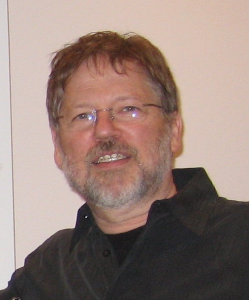
Ted Supalla
Professor, Neurology
Center for Brain Plasticity and Recovery
trs53@georgetown.edu
Education: Ph.D. University of California, San Diego, Psychology, 1982
Current Research: Dr. Ted Supalla’s research centers on the structure and emergence of sign languages around the world. This has included studies of American Sign Language acquisition and processing, and the evolution and structure of homesign, international pidgin sign, and signed languages in other countries. Comparisons between early and modern ASL, and also between other young and well-developed sign languages, can provide an understanding of how languages form and change, and whether the processes of language change for sign languages are the same as those for spoken languages. Dr. Supalla is also interested in the on-line processing of ASL by native signers, including studies of sentence comprehension, sentence reproduction, and short-term memory in signed versus spoken languages, as well as fMRI studies asking what parts of the brain are activated during visual-gestural language and non-linguistic processing.
Peter Turkeltaub

Peter Turkeltaub
Professor, Neurology
Cognitive Recovery Lab
turkeltp@georgetown.edu
Education: M.D., Ph.D. Georgetown University, Neuroscience 2005
Current Research: Peter Turkeltaub is a cognitive neurologist and neuroscientist whose research investigates the brain’s organization for language and other cognitive faculties, how this organization changes in the context of developmental or acquired brain disorders, and how we might enhance recovery. The work is conducted using healthy volunteers and individuals with developmental and acquired language disorders. We use a variety of techniques, primarily TMS, tDCS, fMRI, neuroimaging meta-analysis, and neuropsychology. We aim to expand our methods to include voxel-based lesion analysis, combined tDCS/fMRI, and combined TMS/EEG. A key aim of the laboratory is to develop new treatments for language disorders and translate these treatments to the bedside.
Michael Ullman
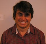
Michael Ullman
Professor, Department of Neuroscience
Brain and Language Lab
michael@georgetown.edu
Education: Ph.D. MIT, 1993
Current Research: Dr. Ullman’s research investigates the neural and computational bases of both first and second language, how language and memory are affected in various disorders (e.g., autism, Tourette syndrome, Specific Language Impairment, Parkinson’s disease, Alzheimer’s disease), and how factors such as sex (male vs. female), handedness (left vs. right), genetic variability, and hormones (e.g., estrogen) affect the neurocognition of language and memory.
Jeff Urbach

Jeff Urbach
Professor, Department of Physics
Dynamics Imaging Laboratory
urbachj@georgetown.edu
Education: Ph.D Stanford, Physics, 1993
Current Research: Dr. Urbach has been actively involved in a broad range of interdisciplinary research and training activities. A physicist with a background in materials science and nonlinear dynamics, he began collaborative work in neuroscience over a decade ago. His recent research has focused on using advanced materials fabrication and live cell imaging technologies to elucidate the role of mechanical and structural cues in axon outgrowth and guidance, with the goal of developing novel strategies to engineer nerve regeneration after injury.
Chandan Vaidya

Chandan Vaidya
Professor, Department of Psychology
The Developmental Cognitive Neuroscience Laboratory
cjv2@georgetown.edu
Education: Ph.D. Syracuse University, 1997; M.S. Syracuse University, 1989; B.A. Bombay University, 1984
Current Research: Dr. Vaidya’s research program is focused upon characterizing the functional neural architecture of adaptive mechanisms during the life span. Adaptive mechanisms promote goal-directed behaviors that allow us to adjust effectively to our environment. Her research focuses on two types of adaptive mechanisms – 1) Processes that require little effort such as learning from environmental regularities without intention or conscious awareness (termed implicit memory and learning); and 2) Processes that are effortful such as voluntary control over thoughts and actions (termed executive control). Her studies investigate how these adaptive mechanisms differ across individuals. Sources of variance under investigation include but are not limited to, genetic functional polymorphisms of catecholamines (e.g., DAT, SERT), metabolic differences (e.g., obesity and its intervention), and psychiatric diagnoses (e.g., Attention Deficit Hyperactivity Disorder, Autism Spectrum Disorders, Schizophrenia).
Dr. Vaidya’s research involves multidisciplinary methods, comprising behavioral, neuropsychological, and structural and functional magnetic resonance imaging in children and adult populations.
Ashley VanMeter
Ashley VanMeter
Director of CFMI; Professor, Department of Neurology
Center for Functional and Molecular Imaging
jwv5@georgetown.edu
Education: Ph.D., Dartmouth College, 1993
Current Research: My research interests are varied but focus on neurodevelopment, reward processing, and neuroimaging methodology. My other area of focus the effects of different environmental influences such as early substance use initiation on the developing. As the director of the neuroimaging center, I’m also involved in a number of other studies.
Stefano Vicini

Stefano Vicini
Professor, Pharmacology & Physiology
Vicini Lab
svicin01@georgetown.edu
Education: Doctorate, University of Torino, Italy, 1979
Current Research: Our studies with Dr. Niaz Sahibzada focus on the major brain nuclei of this circuit that form the dorsal vagal complex in the hindbrain, namely the medial nucleus tractus solitarius (mNTS) and dorsal motor nucleus of the vagus (DMV), indicate a major role of GABA and glutamate signaling from specific local neurons for modulation of vagal output to the stomach.
With our collaborator Dr. Daniel Pak, we hypothesize that impaired homeostatic control may be the cause of Alzheimer’s Disease. We are investigating the effect of specific amyloid precursor protein phosphorylation sites on synaptic plasticity in primary culture and novel knock-in mice.
In collaboration with Dr. Katherine Conant and Dr. Jian young Wu, we are investigating the dysregulation of sharp wave ripples that have a critical role in memory consolidation, and are regulated by parvalbumin inhibitory interneurons ensheathed by perineuronal nets via electrophysiology, and calcium imaging.
Microglial are important players in Alzheimer’s Disease. In collaboration with Dr. William Rebeck, we aim to further characterize morphology and the microglial motility response to ATP in brain slices of APOE knock-in mice, which express the human APOE.
The major goals of the project in collaboration with Dr. Patrick Forcelli are to evaluate with local field potential and patch-clamp recordings from hippocampal neurons the effect on excitatory synaptic transmission of hypoxia-induced seizures and the effect of most common anti-seizure drugs used in infants.
In collaboration with Dr. Mark Burns, we are studying in hippocampal neurons brain slices synaptic adaptation after exposure to head impacts that underlie changes to cognition. We are employing a combination of electrophysiology and calcium imaging and focus on neurons identified to be part of memory engrams with parallel behavioral conditioning.
Tingting Wang

Tingting Wang
Assistant Professor, Pharmacology and Physiology
Wang Lab Website
tw652@georgetown.edu
Education: B.S., Biological Sciences, Tsinghua University; Ph.D., Neurobiology, Duke University
Current Research: The brain is incredibly complex and remarkably malleable in terms of developmental and learning-related plasticity. Homeostatic signaling systems act to stabilize the function of individual nerve cells and neural circuitry, thereby ensuring robust and reproducible brain function and behavior throughout life. My laboratory investigates the molecular mechanisms that underlie the homeostatic control of the nervous system. We are particularly interested in the intercellular signals that convey information between different types of cells during homeostatic plasticity. We use an array of electrophysiological, imaging, molecular and genetic tools in Drosophila melanogaster and mice to address:
- What is the signaling function of glia in synaptic homeostatic plasticity?
- How does dynamic regulation of extracellular matrix (ECM) and adhesion molecules modulate synaptic transmission?
- How do genetic mutations and failed homeostasis contribute to neurological and psychiatric diseases, such as epilepsy and autism spectrum disorders?
Anton Wellstein

Anton Wellstein
Professor, Oncology & Pharmacology
wellstea@georgetown.edu
Education: M.D. J.Gutenberg-University, Mainz, Germany, 1973; Ph.D. J.W. Goethe-University, Frankfurt, Germany, 1985
Current Research: My laboratory has been centered around angiogenesis factors released from tumor cells as molecularly defined therapeutic targets. We have purified a novel heparin-binding polypeptide growth factor (pleiotrophin, PTN) from supernatants of breast cancer cells and cloned the respective genomic and cDNA. The respective protein is secreted from different human tumor cells, is expressed in a number of primary human tumors (breast, prostate and lung cancer and melanoma), and can function as an angiogenesis factor. Since this gene is normally only expressed at appreciable levels during embryonal development, we have been interested in the mechanisms depression during the malignant process and have charactered different promoter and enhancer elements as regulators of the gene. In addition, we have generated different cell lines overexpressing the pleiotrophin gene to obtain defined targets for in vitro and in vivo studies of drugs that inhibit this gene product and have targeted the transcript with ribozymes. Finally, we have generated sufficient amounts of recombinant pleiotrophin protein to characterize signal transduction and clone a candidate receptor cDNA by expression screening.
The laboratory applies numerous different molecular techniques involving DNA, RNA as well as proteins in mammalian and other eukaryotic cells, including extensive cell biology, protein and RNA detection in cells and tissues, gene cloning, regulation and expression studies. Cellular or animal models of development/differentiation and diseases, human tissue or bodily fluid samples from patients are used. Ph.D. students in the laboratory would be able to chose the appropriate technique to answer the question being asked in their research.
Jian-young Wu

Jian-young Wu
Professor, Neuroscience
Laboratory of Cortical Dynamics
wuj@georgetown.edu
Education: M.A., Ph.D, Peking University, 1983, 1986
Current Research: We use voltage-sensitive dyes to visualize cortical neuronal activity and spatiotemporal dynamics of populational neuronal activity. Brain functions are carried out by the activation of a large number of neurons. Spatiotemporal patterns such as oscillations and propagating waves are frequently seen during normal brain functions such as sensory processing, motor planning, thinking, sleeping, and a variety of pathological conditions such as epilepsy and Parkinsons. We study whether wave patterns ( such as spirals and wave compressions) contribute to normal and pathological processes in the cortex. Our ultimate goal is to understand how the functions of the cortex emerge from the organized activity of a large number of neurons.
